Shop by Department
The tunica is a strong, fibrous covering of tissue that covers each testicle. The tunica vaginalis is the exterior layer, while the tunica albuginea is the inner layer. The testicle is separated into sections known as lobules. Seminiferous tubules are small U-shaped tubes found in each lobule. Each testicle has around 800 seminiferous tubules that are tightly coiled. The seminiferous tubules open into the rete testis, which is a network of uncoiled, linked channels. Ducts, or tubes, connect the rete testis to the epididymis, a tightly coiled tube. The epididymis connects to the vas deferens, a long, large duct. A spermatic cord holds each testicle in the scrotum. Each spermatic cord is constructed of thick connective tissue and muscle. It houses the vas deferens, as well as blood vessels, lymph vessels, and nerves.
SKU: LKBI 053/2 13
Categories: Lab Equipment, Biology Lab, Slides
Enter your pin code to check if the product is available
| Specification:- | |
| Shape | Rectangular |
| Material | Glass |
| Thickness | 2-3 mm |
| Usage | Laboratory purpose |
| No. of slides in a set | 1 |
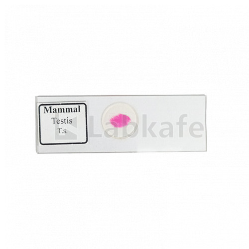
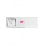
| Sample | Mammal Testis |