Table of contents
- Introduction
- Why have layers of the Human Skin Model become important in a science class?
- Features of Labkafe’s layers of the Human Skin Model
- Where the Human Skin Cross-section model fits best
- Conclusion
- Wants to make your classrooms into demonstration labs.
Introduction
The skin is a complex, multi-layered, and the largest organ in the body, covering the external surface of the human body. It plays a crucial role in regulation, sensory, and defense. There are three layers of the human skin model that have different anatomical and structural functions. Understanding the anatomical structure of the human skin is essential for students of biology, medical science, and bioethicists. To help the students truly grasp the complex concept, Labkafe initiates the layers of the Human Skin model. Cross-section, a hands-on, well-labeled 3D tool that takes a step beyond the textbook diagrams to life. Designed for immense learning, this model makes an illusional diagram visible.
Why have layers of the human Skin Model become important in a science class?
Traditional textbooks in India are often packed with information, but they don’t always make it easy to understand how anatomical structures look or work. Furthermore, for any biology and medical science students, the ability to explain each concept with a realistic 3D layer of the Human Skin model and interact will greatly enhance their knowledge of comprehension, and retention. The 3D model of Human Skin aids in achieving an indispensable result by offering a clear and defined demonstration of the structure. Hereby, filling the conceptual gap between theoretical knowledge and practical understanding. It leads the teachers to promote interactive, discussion-based learning in the biology classroom.
Features of Labkafe’s layers of the Human Skin Model
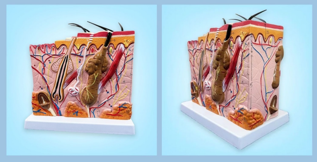
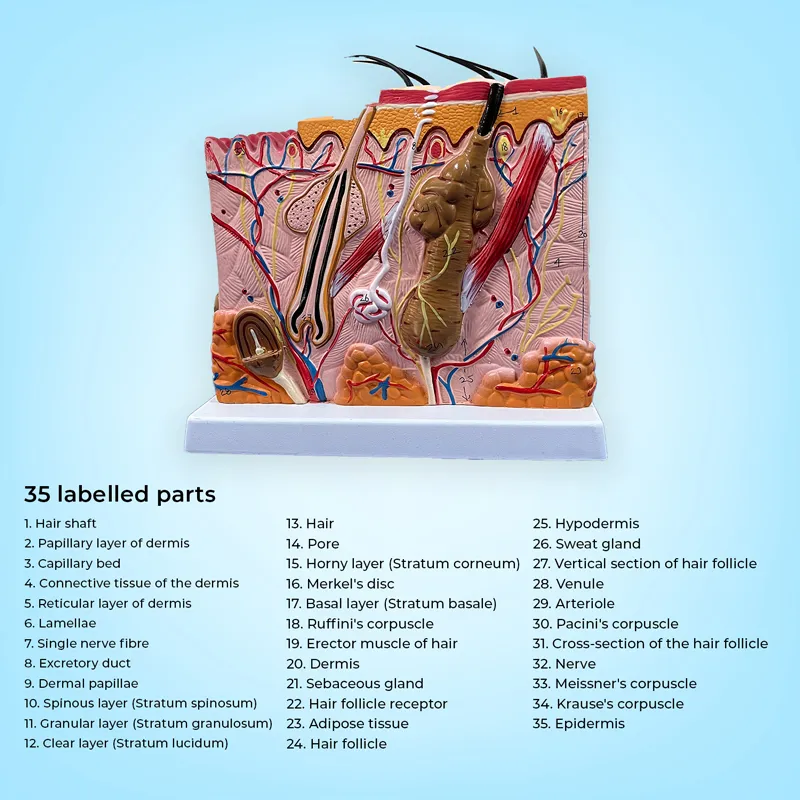
Labkafe’s 3D Human skin cross-section model is developed for academic institutions and teachers’ laboratories. This model provides an enlarged, anatomically accurate cross-sectional model of human skin, allowing the students to explore the structure in a vivid view. Each layer of the epidermis, dermis, and hypodermis is distinguishable and labeled with a scientifically color-coded scheme.
The model is well-defined with 35 labels, making it simple and create visuals perfect for the classrooms. Additionally, it is made up of a high-grade, durable, sturdy wooden frame for easy display, built to withstand repeated handling in a busy laboratory. Every anatomical structure and labeling of hair, glands, and nerve endings is precisely labeled with standard biology and medical-related curriculum. It supports building the foundation and advanced knowledge for any level of academics.
Where the Human Skin Cross-section model fits best
Firstly, the Labkafe’s Human Skin model is a versatile, vivid, and 3D structure that involves NCERT, ICSE, ISE, Medical, UG, as well as PG level students. It helps students to visualize complex biological terms, which aligns with the academic syllabus. It supports advanced anatomical studies in a simple 30×20×25 cm wooden block for nursing, physiotherapy, and aligned health problems. Furthermore, it is an ideal demonstration model for dermatology and a well-defined model for medical students. It becomes an effective tool for demonstrating core skills and allows future professionals to grasp the opportunity to gain hands-on experience through the Labkafe Anatomy Human Skin model.
Conclusion
In a time where experiential learning is critical, Labkafe’s Human Skin Demonstration Model equips educators with a reliable, scientifically accurate tool. Furthermore, it enhances classroom instruction for both primary and higher-level academic laboratories. It turns theoretical concepts into interactive, visual experiences that students can engage with and remember.
Wants to make your classrooms into demonstration labs
Explore Labkafe’s Biology models to access the information about the layers of the human Skin model. Visit our online store to get your Demonstration models on various biological concepts.


Leave a Reply